Chest X-ray in Orlando: Purpose, Procedure, and Risks
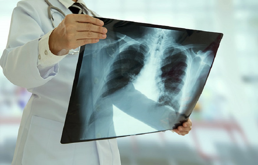
Modern medicine offers patients a wide variety of diagnostic and treatment methods. Today medicine has stepped forward, and doctors do not need to use a scalpel to see the internal organs. Doctors are trying in every possible way to avoid injury, so the inventors have created many new diagnostic devices.
However, some methods were invented many decades ago, but they still have not lost their relevance. Very often, the list of preventive and diagnostic studies includes the X-ray. About 40% of all X-ray examinations are performed to diagnose chest pathologies. This test is necessary for early diagnosis and timely treatment of heart and lung diseases. A chest X-ray is a non-invasive, painless imaging diagnostic method.
Indications for a chest x-ray
A chest X-ray is useful for diagnosing a wide range of diseases. Doctors of almost all specialties appreciate this test. You may be prescribed a chest X-ray by a traumatologist, cardiologist, therapist, neuropathologist, pulmonologist, and other narrowly focused specialists. Doctors of the chest x-ray service in Orlando conduct research to identify pathological processes in the respiratory, cardiovascular, or skeletal systems. Your doctor may also prescribe a follow-up x-ray to check how you are responding to treatment.
You may need a chest x-ray if:
- you complain of pain in the back, between the shoulder blades and chest, especially if you have suffered from a fall or bruise;
- you are the victim of a car accident, which is almost always accompanied by a high injury rate;
- you have a severe persistent cough, shortness of breath, general weakness, weight loss, and high temperature;
- you complain about pain and a feeling of heaviness in the heart, tachycardia with shortness of breath;
- you have the pathological shape of the chest.
What diseases can a chest X-ray reveal?
With the help of high-quality digital equipment, an experienced radiologist without difficulties can identify signs of the following diseases:
- pulmonary tuberculosis;
- pneumonia;
- pleurisy;
- bronchitis;
- asthma;
- lung abscess;
- hydrothorax and pneumothorax;
- heart defects;
- pulmonary embolism;
- diseases of the thoracic spine and ribs;
- tumor.
Are there any risks?
During X-ray examination, the human body is exposed to radiation. Modern clinics use only new digital equipment, which eliminates radiation exposure. When conducting an X-ray of the chest in the X-ray machine, it is possible to adjust the irradiation field for precise aiming at the study area without affecting the rest of the body. So there are no risks of a chest X-ray. This study is also suitable for people of all ages and genders.
How is a chest x-ray done?
The procedure takes place in a specially equipped room. The study is carried out in two projections: in front and side. The doctor asks you to remove all metal objects and leave them in the dressing room. You have to remain still at the command of the radiologist. The doctor will ask you to breathe in or out. The procedure itself lasts less than a minute, after which the radiologist analyzes the data obtained and makes a conclusion.
Other chest studies
To examine the pathologies of chest organs, the health care provider may also recommend fluoroscopy and CT. However, they are prescribed only for the final confirmation of the diagnosis. Most often, to obtain objective data, X-rays are complemented by ultrasound and magnetic resonance imaging. You can read more about MRI of the chest here: https://mricfl.com/s_mri/mri-chest/.

 Treating Severe Burn Wounds with New Methods
  How to Choose the Best Intensive Outpatient Program
  Important Things to Learn About Neurological Physiotherapy Treatment
  Factors to Consider Before Getting a Pelvic Floor Treatment
  Sober Living Homes Helps To Lead A Truly Sober Life After Rehab Treatment
  The Cost of Fertility Treatment: Is It Worth It?
  The Hidden Impact of Micro-Decisions: How Small Choices Shape Big Outcomes with insights from Joe Kiani, Masimo founder
  Teeth Whitening Newport Beach: What Results Can You Expect After One Session?
  NATURAL AND MINDFUL WAYS TO MANAGE NICOTINE CRAVINGS
  How to Choose the Best Products for Hyperpigmentation Based on Your Skin Type